Peripheral Arterial Tonometry (PAT) vs. Photoplethysmography (PPG)
Introduction
Regarding monitoring vascular health and measuring blood flow, two widely used technologies are discussed: Peripheral Arterial Tonometry (PAT) and Photoplethysmography (PPG). Both are non-invasive methods that assess blood volume changes, but they function differently and have distinct applications.
This blog will break down the key differences and how each works.
PAT vs. PPG: Key Differences
The key differences between PAT and PPG are as follows:
|
Parameters
|
PAT
|
PPG
|
|
Technology Type
|
Plethysmographic & Pneumatic
|
Optical-based
|
|
Principle
|
Measures arterial tone via pulsatile pressure changes.
|
Measures blood volume changes via light absorption.
|
| Site of Measurement |
Finger
|
Finger, wrist, ear, forehead, etc.
|
| Primary Use |
- Endothelial function assessment
- Sleep apnea detection - Autonomic and sympathetic nervous system monitoring -Cardiovascular disease monitoring
|
- Heart rate monitoring
- Oxygen saturation (SpO₂) measurement - Blood pressure estimation -Sleep apnea detection
|
|
Influence of Motion
|
Less affected by motion and breathing artifacts.
|
Highly sensitive to motion related artifacts
|
| Clinical Applicatios |
Sleep apnea detection, endothelial function assessment, etc.
|
Heart rate monitoring, fitness tracking, pulse oximetry, etc.
|
Why Choose PPG Over PAT?
- PPG provides non-invasive measurements, requiring no complex devices, making it comfortable and user-friendly for continuous use.
- PPG is cost-effective and offers real-time monitoring.
- PPG is preferred in wearables (smartwatches), fitness trackers, pulse oximeters, and other medical devices. It efficiently measures heart rate and SpO₂ levels, ensuring comfort and seamless health monitoring.
- PPG uses light-based sensors, making it a simpler and more universally adopted method for tracking physiological changes.
- PPG sensors use minimal power, making them ideal for battery-powered devices with long-lasting battery life.
What is Peripheral Arterial Tonometry (PAT)?
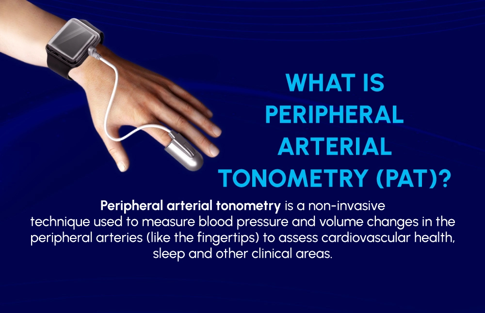
PAT technology is a plethysmographic-based, non-invasive method designed to measure pulsatile volume changes in peripheral arteries, particularly at the fingertip. It measures the blood flow in the small arteries by detecting pressure changes. This measurement helps in the assessment of vascular tone and can also aid in diagnosing conditions like sleep apnea.
It applies gentle pressure to the fingertip, tracking how the arteries expand and contract with each heartbeat. This helps monitor the tone (tightness or relaxation) of the arteries and provides insights into vascular health and diagnosing sleep apnea.
How does PAT work?
PAT technology uses a simple finger probe (biosensor) that measures finger arterial pulsatile volume changes by collecting the PAT signal.
- The finger probe is placed at the distal phalanx (fingertip) as this location ensures accurate detection of vascular tone changes.
- The finger probe's pressure biosensor applies a uniform sub-diastolic covering the mid and distal phalanges of the finger. This pressure helps relieve arterial wall tension and prevents venous blood pooling. Moreover, the PAT probe ensures it clamps to the finger without dislodging.
- As blood flows through the arteries in the fingertip, the biosensor detects minute changes in arterial volume caused by the blood pulses, which is essentially the PAT signal.
- These volume changes provide real-time insights into vascular function and arterial tone.
- The collected PAT signal undergoes advanced processing, and the resulting data is then processed using digital signal processing algorithms for further clinical assessment.
What is Photoplethysmography (PPG)?
PPG is also a non-invasive method, but it uses light to measure blood flow changes in the microvascular tissue. It is commonly found in wearable devices, pulse oximeters, and fitness trackers.
How does PPG work?
PPG operates through the following steps:
- The LED or light source emits light at two specific wavelengths, typically red and infrared.
- The light penetrates the skin and interacts with the blood in the microvascular bed, where it is either absorbed or reflected. As blood volume fluctuates with each heartbeat, the amount of absorbed or reflected light changes.
- A photodetector captures the light that is transmitted through the tissue or reflected from the skin.
- The captured light variations are converted into electrical signals by a photosensitive element.
- These electrical signals are processed and analyzed by a connected application or device.
- The application or device interprets the processed data and displays metrics such as heart rate, blood oxygen levels, apneic events, sleep stage, etc., derived from the PPG signal.
Conclusion
PAT and PPG play vital roles in non-invasive health monitoring but serve different purposes. Choosing the right technology depends on the specific application for clinical use or everyday health tracking.


